| Gypsy moth larvae |
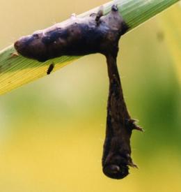 |
| In the late stages of LdMNPV infection, gypsy moth larvae climb to treetops and die. Retrieved from [http://www.nature.com/news/2011/110908/full/news.2011.526.html] |
Although a great diversity of parasite-induced changes in host behaviour has been documented, the physiological mechanisms utilized by these parasites are not well known. Current research has looked primarily at parasite-induced changes to biogenic amine systems. In many animals, biogenic amine systems control a variety of functions including locomotion, feeding, predator avoidance behaviours, and motivation. It is difficult to clearly define pathways and mechanisms of parasitic manipulation in these systems due to the complex range of behaviours affected by even a single transmitter[1]. Nonetheless, changed levels of neurotransmitters such as serotonin, dopamine, and octopamine (an invertebrate neuromodulator) are correlated with abnormal behaviour in infected animals[2]. In addition to modifying biogenic amine systems, some parasites target specific areas and circuitry of their host organism’s central nervous system to affect a particular behaviour[3]. Finally, some studies are using modern genetic and proteomic techniques to look at the way parasites may cause changes through altering their hosts' proteomic profiles[4].
Biogenic Amine Systems
Dopamine
Dopamine in fish is correlated with a wide range of functions, including social behaviour[5], endocrine system regulation[6], and locomotion[7]. This system is implicated in the behavioural change observed in California killifish (Fundulus parvipinnis). The trematode Euhaplorchus californiensis infects killifish, its intermediate host, forming cysts in their brains in which the parasite matures. The parasite must be transmitted to birds, their definitive hosts in which they reproduce. Infected individuals display jerking, contorting, and shimmying behaviours that result in their becoming 30-times more likely to be eaten by birds[8]. Using high-performance liquid chromatography, it was found that experimentally infected killifish had a cyst density-dependent increase in hippocampal serotonin when unstressed[[(bibcite example9))]. They also showed a decrease in serotonin and increase in dopamine in the Raphe nuclei[9]. A follow-up study looking at naturally infected killifish populations found that individuals harboured a higher density of cysts, possibly no significant changes to hippocampal serotonin levels, and a density dependent decrease in dopamine in the Raphe nuclei[10]. The disparity between these two studies suggest that parasitic effects on killifish monoamine levels may be time-dependent[10]. While dopamine and serotonin both correlate to motivation and locomotion, the exact chain of events leading to behavioural alteration is still unknown in this system.
In rodents, locomotion and exploration induced by novelty, stress, or drugs like amphetamine involve dopamine activation, with high exploration being linked to higher dopamine release and numbers of high-affinity D2 receptors in the striatum[11]. When infected with the protozoan parasite Toxoplasma gondii, which forms cysts in the brain, rats and mice exhibit changes to their locomotive behaviour. In open field tests and holeboard tests (testing exploratory behaviour and locomotion), Toxoplasma infection in both sexes decreased locomotion in open field, and in females increased exploration in the holeboard test[12]. Injection of GBR12909, a selective dopamine uptake inhibitor, suppressed exploration in holeboard test in infected males but increased it in infected females[12].
Immunohistochemical assays of the brains of infected female mice have shown that Toxoplasma tissue cysts had high concentrations of dopamine, and that these cysts were more abundant in the amygdala and the nucleus accumbens, which have high concentrations of dopaminergic neurons and are part of the circuits for reward, motivation, fear, and movement[13]. Cultures of Toxoplasma infected neurons had about 300% greater dopamine concentrations as well as dopamine release compared to uninfected cultures, and increased infection intensity had greater effects[[13]. A possible explanation for the increased dopamine metabolism is the heightened production of the enzyme tyrosine hydroxylase, the rate-limiting enzyme in dopamine synthesis which catalyzes the conversion of tyrosine into L-DOPA[[14]. The T. Gondii genome encodes a tyrosine hydroxylase with a unique amino terminal sequence. Staining with antibodies specific to this viral enzyme variant showed high expression specific to encysted neurons[[(bibcite example13))].
Serotonin
| Serotonin in Amphipod Behavioural Change |
 |
| Changes in brain serotonin immunoreactivity in Gammarus pulex infected with P. laevis. Retrieved from [http://jeb.biologists.org/content/216/1/134/F1.expansion.html] |
Behavioural changes in amphipod crustaceans infected with acanthocephalan parasites correlate to changes in serotonin levels. Amphipods are normally photophobic, but infection with certain species of acanthocephalans will cause behaviours such as an attraction to light (photophilia, or positive phototaxis) or an attraction to the water surface (positive geotaxis). The relationship between this behavioural alteration and increased trophic transmission may not be as straightforward as previously hypothesized: while infected amphipods are more likely to be eaten, predation risk of infected individuals did not vary with magnitude of phototaxis (tested by performing trials under different light intensities)[15]. Uninfected amphipods with pharmacologically induced phototaxis were also not likely to be eaten than uninfected individuals given a neutral injection. This implies that, at least in infection of Gammarus pulex by Pomphorynchus tereticolli, that phototaxis is a consequence of infection and not a directly manipulated trait for increased parasite fitness[15].
Different acanthocephalan species may induce different behaviours in their hosts, and the role of serotonin may differ in each system. Gammarus pulex, a freshwater amphipod, may be infected by two species that cause photophilia (Pomphorynchus tereticolli, Pomphorynchus laevis) and one that causes positive geotaxis (Polymorphus minutus)[16]. Serotonin is specific to phototaxis in this species. The brains of amphipods infected with a phototaxis-inducing species showed significantly higher serotonin immunoreactivity compared to control, whereas those infected with P. minutus did not[16]. Additionally, injection of serotonin into uninfected individuals induced phototaxis comparable to those of infected amphipods but no geotaxis[16].
Infection of Gammarus lacustris by Polymorphus paradoxus induces photophilia and also a disturbed escape response, where the infected animal skims along the water's surface until it finds a solid object to which it clings in a flexed posture. The amphipod may remain in its flexed posture for up to 4 hours before it relaxes, but may continue to grasp the object for a further period of time[17]. Injection of octopamine into infected G. lacustris suppressed the clinging and serotonin induced it in uninfected animals, both in a dose-dependent manner, and the duration of both pharmacological effects were brief[17]. Injections of octopamine, dopamine, norepinephrine, and GABA all failed to elicit the clinging behaviour, showing that this is serotonin dependent[17].
A third amphipod species, Echinogammarus marinus exhibits increased geotaxis and phototaxis when infected with acanthocephalans. Exposure of uninfected individuals to serotonin in the water for over a week led to phototaxis and geotaxis, with the magnitude again being dose-dependent[18]. Exposure to fluoxetine, an SSRI, also significantly increased both behaviours. Carbamazepine and diclofenac, two anti-depressants that do not work via serotonin, had no significant impact[18].
Octopamine
| Cotesia congregata cocoons |
 |
| After the wasp larvae emerge from the hornworm caterpillar, they spin cocoons on the body of their quiescent host. Retrieved from [http://www.discoverlife.org/mp/20p?see=I_JPS1&res=640] |
The parasitoid wasp Cotesia congregata lays its eggs inside the tobacco hornworm Manduca sexta. The larvae develop within the hemocoel, feeding on nutrients in the hemolymph but leaving the internal organs unharmed, and emerge through the hornworm's cuticle when they have moult to the third instar[19]. About 8-12 hours before the wasps emerge, the hornworm stops feeding and crawling. It lives for 2 more weeks in this immobile state, during which the wasps spin their cocoon on the hornworm[19]. This behavioural manipulation is beneficial to the wasps because unparasitized hornworms will eat the cocoons if left unsuppressed, and host mortality do not appear to be higher than that of unparasitized individuals[20].
Studies of this parasite-host system found correlations between hornworm behaviour changes and octopamine, a monoamine with functions in M. sexta such as modulating learning[21] and the flight central pattern generation[22]. Octopamine content in the hornworm's central nervous system increased at the same time as the cessation in feeding and locomotion, an effect that is not due to inactivity since food deprivation and moult sleep, a comparable inactive state in unparasitized hornworms do not increase octopamine[19].
The feeding suppression appears to stem from disruptions to the frontal ganglion of the hornworm by octopamine[23]. Operational removal of it leads to decreased efficiency for ingesting food and decreased the number of feeding bouts a hornworm will make[23]. Electrophysiological recordings showed weakened peristalsis in the esophagus and absent or weak and infrequent spiking in the frontal ganglion after wasp emergence[23]. This electrical disruption can also be achieved by application of hemolymph from post-emergence wasp larvae or octopamine at the concentrations of post-emergence wasp hemolymph to the frontal ganglion[23]. The position of octopamine in feeding suppression is strengthened by the fact that co-application of an octopamine blocker (phentolamine or mianserin) with wasp hemolymph prevented this disruption[23].
Neuroanatomical Targets
Jewel Wasp
| Parasite tales: The jewel wasp's zombie slave |
| Ecology of jewel wasp and cockroach, presented by Carl Zimmer. |
The jewel wasp (Ampulex compressa) is a parasitoid that attacks the cockroach (Periplaneta americana). It stings its victim twice. The first, delivered to the thoracic ganglion, temporarily paralyzes the front legs in order to facilitate the second sting. The second sting induces hypokinesia: the cockroach loses the drive to initiate movement and has dampened escape responses, allowing the wasp to lead the cockroach to its burrow, lay an egg on it, and seal up the nest. When the wasp larva hatches, it feeds on the cockroach.
Pharmacological depletion of dopamine, serotonin, octopamine, and tyramine by injecting reserpine (an inhibitor of vesicular monoamine transporter) into cockroaches results in hypokinetic behaviours similar to those of stung animals[24]. However, analysis of the amine profile of stung cockroaches using high-performance liquid chromatography did not find differences in the levels of the four transmitters of note, indicating that wasp venom does not work via those pathways [24]. On the other hand, opioid antagonists, most prominently Nor-Bni (antagonist for κ-opioid receptor), prevented hypokinesia if injected prior to envenomation. [25]
The jewel wasp has been shown to actively search out and target specific ganglia of cockroach CNS. The head sting, which induces hypokinesia, is delivered to the supra-esophageal ganglion and sub-esophageal ganglion (SEG)[3]. Although the role of the supra-esophageal ganglion remains unclear, the SEG is important for escape behaviour and autonomous movement[3]. Injection of procaine, a sodium-channel blocker, into the SEG decreases autonomous motion[3]. Electrode recordings show that spontaneous spiking activity in the SEG decreases with sting, and that stimuli that normally induce escape behaviour (which is impaired in hypokinetic cockroaches) evoke fewer spikes[3].
Toxoplasma gondii
Toxoplasma gondii infection in rodents causes attraction to cat odours, increasing the likelihood of predation by cats and therefore the transmission of the parasite to cats, the definitive host in which it reproduces[26]. Toxoplasma forms cysts in the brains of rats, which occur in low density in the cerebellum, compact myelinated fibre tracts, commissures, and subcortical structures bounded by myelinated axons, suggesting that cyst are blocked from areas made less accessible by white matter[26]. Cysts are found more densely in cerebral cortical areas than subcortical except for the amygdala and tegmentum, which are highly infested[26]. Although Toxoplasma, like most microscopic parasites are less specific in neuroanatomical targets, there is evidence that the high infection of amygdala facilitates behavioural alteration. Using c-Fos (an immediate early gene) count as a proxy for neural activity, infected and uninfected rats were exposed to either cat urine or the scent of a female rat in estrus, then sacrificed for examination of brain activation[27]. Uninfected rats exposed to cat odour showed high activity in the dorsomedial part of the ventromedial hypothalamus, an area associated with defensive response[27]. Uninfected rats exposed to estrus and infected rats exposed to cat odour both showed increased activation of the posterodorsal medial amygdala, which is associated with reproduction[27]. These studies suggest that Toxoplasma may manipulate neural circuitry to effect behavioural alteration in rats.
Proteomic Mechanisms
LdMNPV
The caterpillar of European gypsy moth, Lymantria dispar, normally hides in bark crevices or soil during daytime to avoid predation and emerge to feed on leaves at night[4]. Following infection by the baculovirus Lymantria dispar nucleopolyhedrovirus (LdMNPV), the caterpillar climbs to the top of its tree, dies, and liquefies, thereupon releasing viral particles[4]. The baculovirus gene ecdysteroid uridine 5'-diphosphate (UDP)-glucosylstransferase (egt) encodes the EGT enzyme, which inactivates the caterpillar molting enzyme 20-hydroxyecdysone (20E)[4]. Experimental infection of caterpillars with engineered egt knock-out strains of LdMNPV died at low elevations in the late stages of infection whereas a strain engineered for rescue of the egt induced death at the same high elevations as wild type virus, demonstrating that this gene is key to manipulating the climbing behaviour of gypsy moth larvae[4].
Rabies
Rabies is a viral disease in mammals with a wide list of symptoms including weakness, anxiety, confusion, insomnia, agitation, hydrophobia, and eventually death[28]. Several recent studies have used proteomics to help unravel the mechanisms of this disease, investigating the workings of several strains of this virus using techniques including 2D electrophoresis, immunohistochemistry, and mass spectroscopy.
A study using mice found that the wild-type SHBRV (silver-haired bat rabies virus) and attenuated B2C strains altered the expression of over 55 proteins combined[28]. The SHBRV strain up-regulated genes coding for proton pumps and Na+/K+ pumps and down-regulated Ca2+ pumps and SNAREs such as α-SNAP, syntaxin-18, and pallidin[28]. Dysregulation of neuronal ion concentrations resulting from changes to ion channel expression may be the molecular cause of the seizures observed in mice infected with this strain[28]. The down-regulation of SNARE proteins led to accumulation of vesicles in the axon terminal, observable under electron microscopy, and therefore to a disruption in neurotransmitter release and uptake that would result in a slew of CNS dysfunctions[28]. In contrast, B2C infected mice notable showed down-regulation of proton pumps and up-regulation of APAF1, a marker for apoptosis that is involved in caspase activation[28]. Down-regulating protron pumps trigger cellular acidosis, which would also lead to apoptosis[28]. These molecular changes are consistent with the edema, ischemic injury and neuronal damage as a result of inflammation, necrosis, and apoptosis observed in B2C mice[28].
Another study looked at the challenge standard virus and the Pasteur virus in a mouse neuroblastoma line[29]. 40 proteins out of the 64 differentially expressed spots were identified, with almost half being cytoskeletal proteins, including overexpression of three vimentin proteins, related to intermediate filaments, and new induction of six more[29]. Two proteins related to actin filaments were also underexpressed, and in particular profilin I is involved in vesicle recycling[29].
Intro looks good! Concise and clear. I'm definitely going to be linking this to my page :)
Definitely! I think I've got some good supplementary material for your section, since we do have a lot of organisms in common. Yours look awesome already, btw! Love the zombie media!
This is why I love Neuroscience. Thanks for the excellence and collegiality from both of you. I know this might be construed as a challenging project but think of how amazing your work will be and how many people will be able to see how well you can write, edit and independently research! I have loved having both of you as students this past year. Bill
Thanks Dr. Ju! I thought this was pretty fun, and it got me exploring and talking about a topic I might not have look seriously at otherwise. I'm gonna confess, at the eleventh hour here, that I'm having a bit of an existential crisis about my page because there is actually a lot of bits and pieces here and there that I couldn't fit into this.
Your page looks great so far!
I'm really interested to read your section on serotonin. My page is about the serotonin theory of Autism, so it would be interesting if we could link our pages!
Thank you, and I'd love to! It'll be a nice strange surprise for whoever goes from your page to mine, haha!