| Mindfulness State through Meditation |
 |
| From:http://fusionwellness.ca/?attachment_id=1575 |
The neurological aspects of the mindfulness state, non-judgmental physical and emotional self-awareness have become widely studied over the past years, as they are associated with stress and depression reduction[2]. Specifically, researchers found neuroplasticity in various areas in the brain that are associated with emotion regulation, during and after the mindfulness practices of the patients[1],[22]. There are various approaches to reach the mindfulness states, in which each one of them affects the brain differently.
The 4 Common Approaches[1][2][3]:
- Mindfulness-Based Stress Reduction
- Mindfulness-Based Cognitive Therapy
- Mind-Body Training
- Mindful Attention Training
These approaches became possible interventions for patients that are suffering from stress-related psychological disorders such as anxiety disorders and depression [2].
|
Table of Contents
|
1. Mindfulness-Based Stress Reduction
1.1 DMN
 |
| (A and B) shows activation in MPFC and PCC. (C and D) the percentage change in activation of mPFC and PCC in: Awareness (green), Loving-Kindness (red), and Concentration (blue) Meditation. Figure from [10] |
The most extensively researched technique is called, the ‘Mindfulness-based stress reduction’ method (MBSR), which combines many practices of sitting meditation and yoga. The most observable stress reduction effect is deactivation in main areas of the default mode network (DMN), a mind-wondering state that is associated with depression[11]. DMN is a self-referential processing that comprises of medial prefrontal cortex (mPFC), posterior cingulate cortices (PCC), hippocampus, and amygdala[11]. Furthermore, there is also deactivation in the mPFC and PCC of individuals who performed MBSR for 8 weeks[10]. Reductions in mPFC specifically increase the present moment self-awareness, which is beneficial for individuals to prevent their undesirable self-reflection eleboration[23].
A study on gamma power also supports this concept as the EEG gamma power especially in the frontal and the midline structures are positively correlated with the DMN activity level[11]. It is found that there is a lower gamma level in the frontal and midline areas in meditators, represents a decrease in DMN activities level[11]. Furthermore, there is an increase in the connectivity of the DMN with task-positive regions and self-control regions in the brain of MBRS individuals[11]. Consequently, these regions have roles in regulating attention and reducing negative interpretation that is important for individuals to regulate their perceptions in stressful situations. However, DMN is on of many research areas that are full with contraries as many researches have distinctive results due to different approaches of mindfulness practices.
1.2 SRN
In contrast from the above DMN concepts, a research on the self-referential network (SRN) that also focuses on similar regions as the DMN point out contrary results. By using fMRI to measure BOLD level in Social Anxiety Disorder patients (SAD), it is revealed that MBSR increases activations in the PCC and DMPFC areas, which will down regulate unconstructive feelings in the patients[10]. It is further specified that functions of PCC and DMPFC not only includes self-referential processing, but as well as emotion awareness and mood regulation. Therefore by increasing the DMN activation will aid SAD patients to decrease their ‘distorted self-evaluation’[10].
1.3 Gray Matter Concentration
 |
| There is a significant increase in the gray matter density of PCC in the MBSR individuals. Figure from [4] |
There is an increase in a gray matter concentration in the left Hippocampus of individuals that practiced MBSR for 8 weeks[4]. As hippocampus generally involves with emotion regulation, neuroplasticity in this area may improve emotion and stress control. Moreover, some researches have found that a reduction in the hippocampus volume can be linked to the development of stress-related disorders such as post-traumatic stress disorder and major depression[4]. There are also significant increase in gray matter concentration in temporo-parietal junction (TPJ), PCC, and cerebellum; which all of these areas contribute to self-referential processing, self-perception, mood regulation, social cognition, and compassion [4],[14],[15].
2. Mindfulness-Based Cognitive Therapy
2.1 Frontal Brain Asymmetry
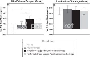 |
| Rumination group has higher EGG alpha activity . Figure from [5] |
Researchers have been associating the frontal alpha asymmetry with depression since the past decade. The earlier studies in the 20th century associate the left frontal lobe with positive emotions, whereas the right lobe relates to anxiety and avoidant behavior[16]. Moreover, they discovered that depressed people commonly have higher EEG alpha activity level in the right frontal lobe relative to the left lobe, in the non-depressed people[17].
However, in more recent researches starting from few years ago suggest that higher EEG activity in the right frontal lobe can be associated with motivation, which can be interpreted in either way of positive or negative emotion (enthusiasm or anger)[18]. It is shown in a MBCT study on depressed women that mindfulness practices promote the frontal asymmetry by increasing the right frontal EEG alpha by greater amount than the left EEG[5]. This study explains further that the MBCT approach maybe an intervention to prevent relapse in major depression[15].
2.2 Pain
Pain is one of the factors that can result in stress and depression. According to many researches, meditation engaged in various brain mechanisms that may reduce the activation of the contra lateral primary somatosensory cortex in the nociceptive area, by activating ACC, AI, and OFC, or the three executive regions of attention control[13]. ACC and anterior insula (AI) regulate the cognition of nociceptive processing, whereas orbitofrontal cortex (OFC) controls the contextual evaluation[13]. An experiment conducted by F. Zeidan and others consisted of 2 conditions (heat and neutral), particularly they applied thermal stimuli to trigger pain and unpleasantness in the participants[7]. By MRI scanning the participants during the mindfulness state, it is found that pain intensity declines with the activation of ACC and AI, whereas unpleasantness level due to pain decreases accordingly to the activation of OFC[7]. Only after 4 days of the experiment, there is a substantial reduction in the pain intensity as well as the unpleasantness due to pain in the meditator group compared to the control group, 40% and 57% accordingly[7].
3. Mind-Body Training
3.1 White Matter Efficiency
 |
| After 4 weeks of MBT, FA increases, AD and RD decrease. Figure from [9] |
By practicing Mind-Body training, which is to release negative thoughts and to be aware of all sensations while meditating, measurement of fractional anisotropy (FA) increases around that anterior cingulate cortex[6]. FA is a value that reflects white matter myelination, and it increases when both radial diffusivity (RD) and axial diffusivity (AD) decreases; however, FA value can also increase when only RD[19]. An experiment done by Yi-Yuan Tang and others discovered that the white matter efficiency of ACC increases as both AD and RD decreases or only RD decreases after 4 weeks of MBT practices; consequently this results in an increase of the self-regulation network[6]. Furthermore, this experiment also depicts that RD is an important factor to increase the white matter efficiency, and there are results from other experiments that RD has a negative correlation with myelination[6],[8]. Increase in the white matter efficiency does not only enhance the self-regulation network but as well as conflict solving ability; these are the aspects that are needed under stressful situations[19]. ACC is a critical area for emotion regulation; therefore it is being affected in almost every mindfulness methods[9].
4. Mindful Attention Training
4.1 Amygdala
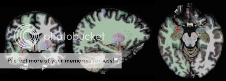 |
| The red crosses on the blue areas marked the amygdala. Coronal, sagittal, and horizontal views. Figure from [3] |
Mindfulness Attention Training is different from the other practices as it is based on 3 meditation focuses, which are breathing, thoughts, and awareness[24]. After the MAT training, there is an activity reduction in the right amygdala, which is the area associated with motivation, emotion and behavior[3],[20]. Hyperactivity in the amygdala may result in emotional disturbances such as stress an anxiety[20],[21]. Furthermore, being stressed over a certain period of time may correspondingly lead to an increase in the amygdala activity[21]. A research done by Desbordes and others indicated that participants that are under the MAT training for 8 weeks have amygdala activity reduction after they were shown with emotional images. Thus, this suggests that mindfulness effects during meditating might be able to unconsciously transfer to the normal non-mindfulness state[3].
5. Long-term Effects of Meditation
Individuals with long meditation experience have different reaction and emotion regulation from none-meditative individuals. A research done by Sobolewskia used visual event-related potential (ERP) test to measure brain response according to visual stimuli. Specifically, higher positive ERP value reflects greater intensity in the emotion responses[25]. Experienced meditators were compared with the control group, in which the meditator must have at least 5 years of at least 5-hours/week meditations. After meditators were shown with negative pictures, their ERP remained constant especially over the frontal cortical region; this reflects that meditators can impede arousals that are caused by negative stimulus[25]. Hence, this suggests that maintaining mindfulness state might be able to effectively inhibit negative thoughts about self and surroundings that possibly lead to stress and anxiety. Lastly, it has been analyzed that one of a possible reasons meditation or mindfulness practices can effectively reduce stress is because meditation has a core concept that highly emphasis “detachment”, or the state of letting go all worry and pain[26].
| [TEDxCollegeHill] Mindfulness Starts With the Body:Catherine Kerr |
| Answering many interesting questions related to meditation, such as why we have to focus on our breathing. |
Thanks for putting this up so quickly! Look forward to reading what you have to post and I've greatly enjoyed having you as a student and trying to answer some of your questions this semester. Best wishes, Bill
This sounds like a very interesting topic, I cant wait to read more! For the pictures/ figures, I would suggest adding captions to them.
Interesting Topic!!! On a related note, there is an old paper that suggested that mediation can reduce glutamatergic transmission. Since stress enhances glutamatergic transmission and the excess glutamate activates pathways that harm the cells, mediation can actually reverse the effect of stress by normalizing the glutamate transmission to normal and prevent the activation of harmful pathways.
Here is the abstract of the paper: http://www.ncbi.nlm.nih.gov/pubmed/11958969
hey great topic! it is related to mine as well (which is meditation-induced hallucinations) so I'll be linking your page to mine :) also the last section, the last sentence cuts off without an end to it
Great neurowiki!
Here's a suggestion for an external link: Mindfulness Starts With the Body: A View from the Brain - http://www.youtube.com/watch?v=AGnGRgyLwMs it's a TED talk that elaborates on the research that you have put together.
Great topic! In depth research! but i had trouble viewing images that you inserted between sections. Other than that i really enjoyed reading this wiki!
I thought of doing this topic myself as I have practiced meditation for many years and it really helps me deal with stress. I really liked the section on the amygdala and will be linking my page to yours - as I talk about the effects of music on the amygdala. Good work. The only problem is that your images aren't showing up for me either.
You've chosen a very interesting topic that's recently been getting a lot of attention. I think you've done a wonderful job creating this summary. I noticed a small error in 2.1, I think you may have meant "researchers" for the first word?
I especially found your section about brain lateralization interesting. A lot of this early work was done by Davidson et al. (at Wisconsin-Madison I think?) who conducted the first lateralizationg studies with EEG. I know he subsequently did some work with the Dalai Lama himself and other experienced monks and meditators, showing that they had even greater left frontal lobe activity. Here's a paper you may want to review: http://www.ncbi.nlm.nih.gov/pmc/articles/PMC2267490/ if you're interested. Another fascinating look at the topic is to hear it from a monk directly (and a converted scientist-to-monk at that!). Matthieu Ricard presents some fascinating results and advice in this TED talk: http://www.youtube.com/watch?v=vbLEf4HR74E
It's not directly relevant to your topic, but do check it out for interest if you have time!
Aim~Good job with the work! You taught me so much about mindfulness and how to mediate stress.
Just a heads up that your last sentence in "Long term effect meditation" section is cut off. Also, you might want to add a title to your figures, and for the figure in "Frontal brain asymmetry"section, i think the picture is better if it's larger, so we can see the axis better.
You did a wonderful work!
Nicely done, its put well and I dont see anything wrong. The topic is very interesting, and i found it fascinating. :)